Author Topic: Correlating Images and Point Analyses in PFE  (Read 6707 times)

John Donovan

  • Administrator
  • Emeritus
  • *****
  • Posts: 3304
  • Other duties as assigned...
    • Probe Software
Correlating Images and Point Analyses in PFE
« on: December 31, 2013, 06:11:58 PM »
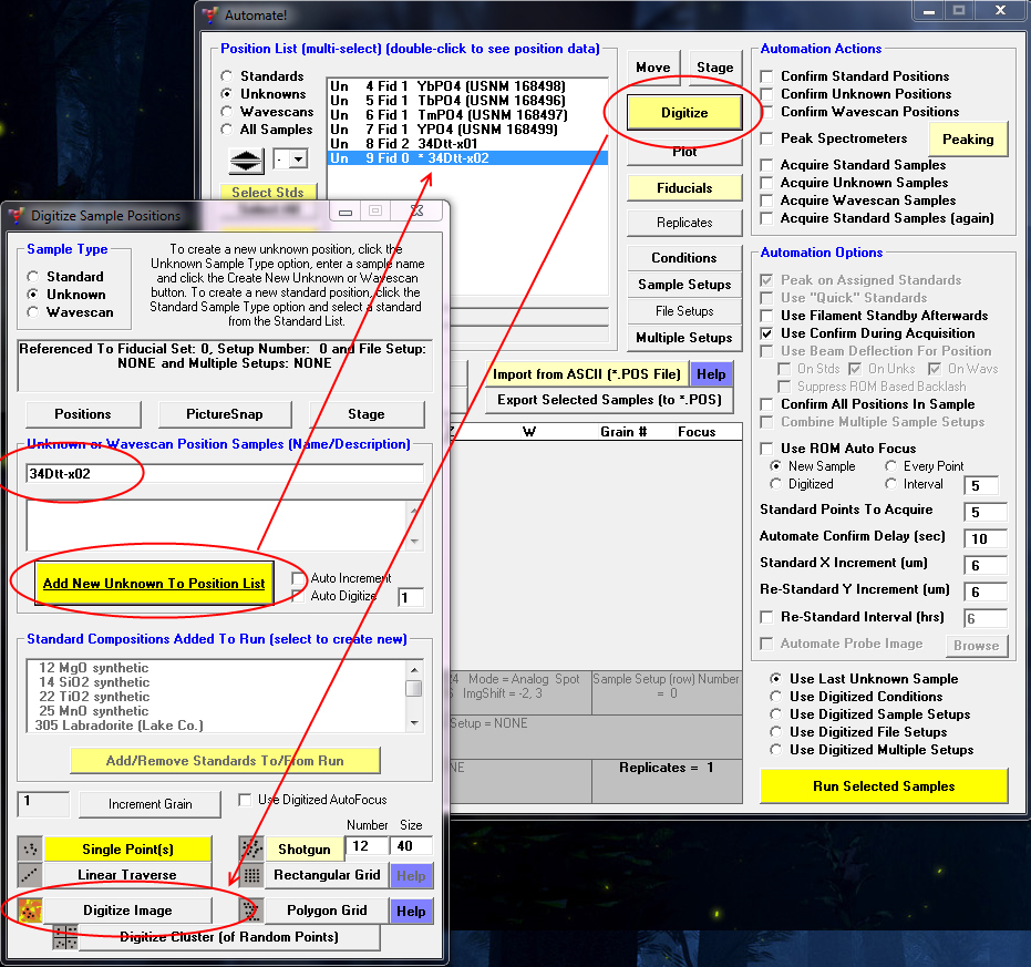
In Probe for EPMA it is easy to acquire BSE, SE, CL, etc. images and also acquire manual or automated point analyses. It is also easy to acquire a BSE, SE, CL, etc image and use the mouse to select point analysis positions on the image as seen here by utilizing the Digitize Image feature accessible from the Digitize window from the Automate! window:


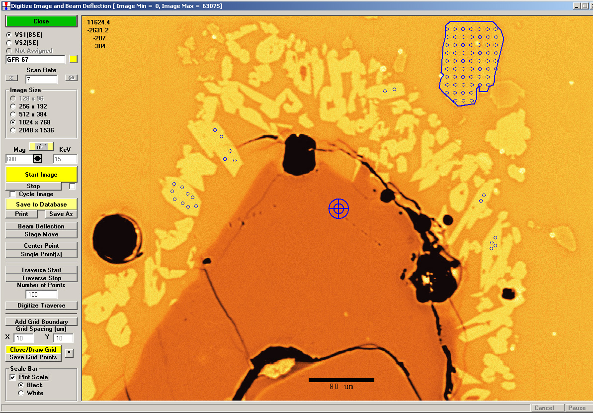
This allows us to easily overlay our point analyses accurately over our analog signal image (BSE, SE, CL, etc) with just a mouse click using the Plot Stage Positions | All Positions option as seen here outlined in red:


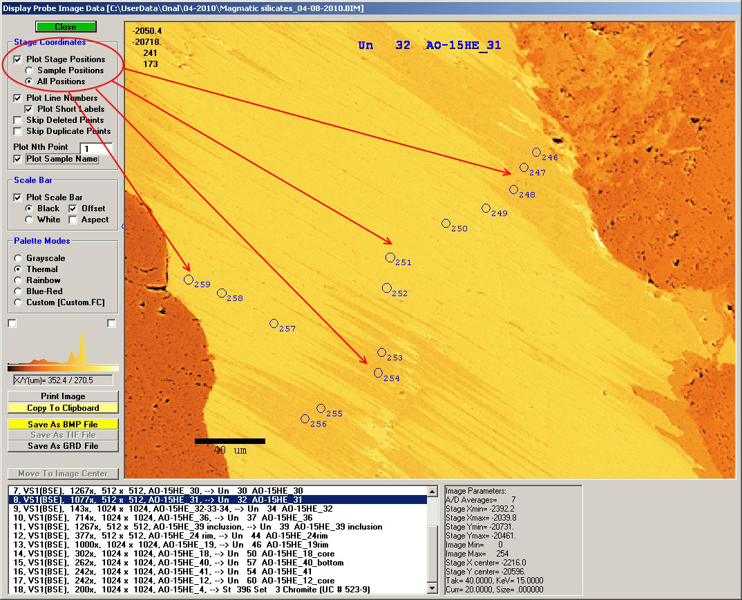
So while one can display any analysis positions within the field of view of any image, sometimes we want to only display the analysis positions of a specific sample as seen here:


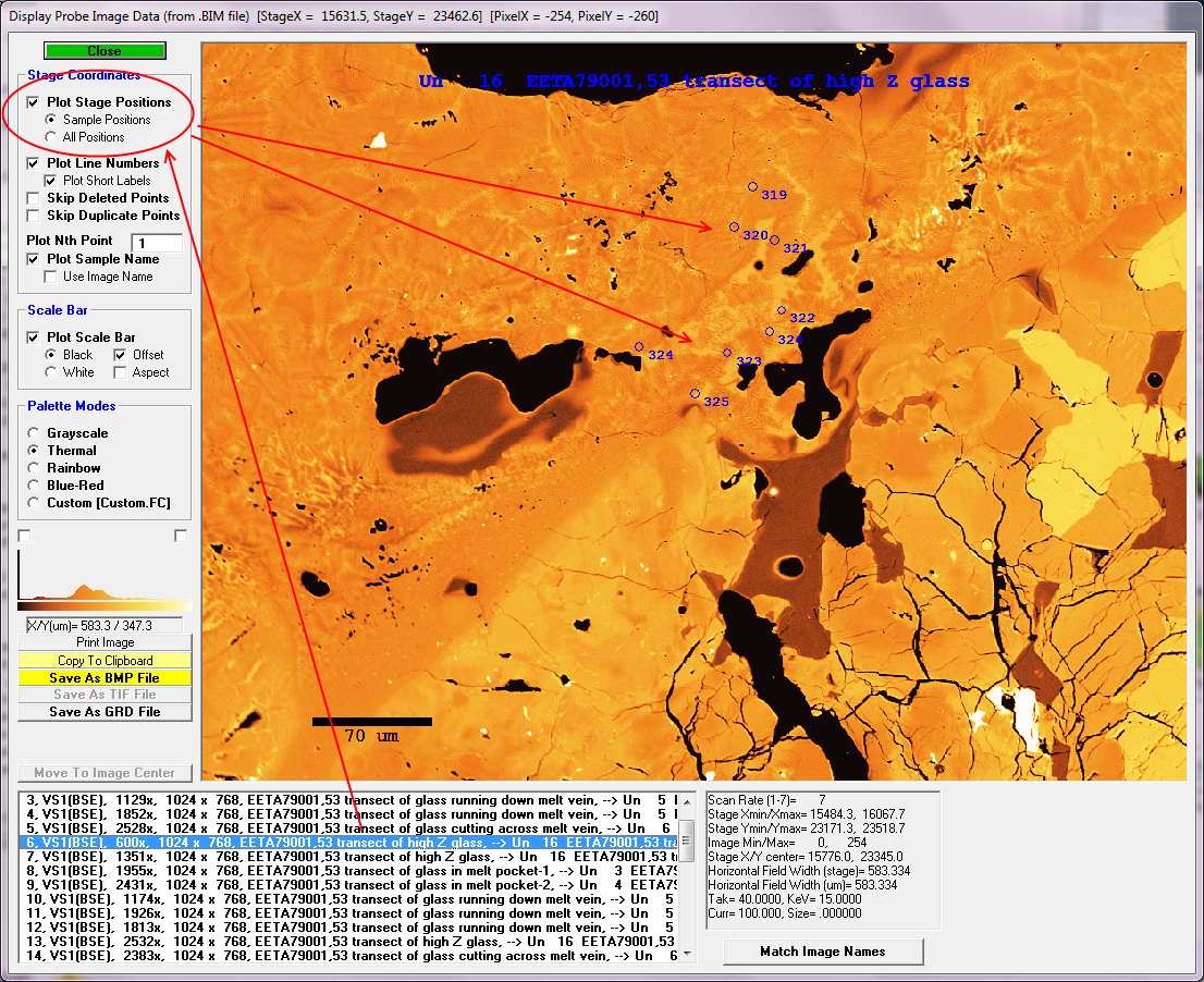
The easiest way to accomplish this is to enter the same name for the image and the sample as we acquire images and specify point analysis positions regardless of whether the images are acquires manually, automatically or using the Digitize Image feature as described above as as seen here:


Once the point analyses have all been acquired manually or automatically, we can go to the Run | Display, Annotate and Export Analog Signal Images menu and click the Match Sample Names button as seen here:


The program will search through the probe run and locate any samples that match the image names and specify a "pointer" (the --> arrow in the image description) that indicates the point analyses that correspond to the image.

Now the Plot Stage Positions | Sample Positions option will display only those point analyses that are contained in the specified sample.

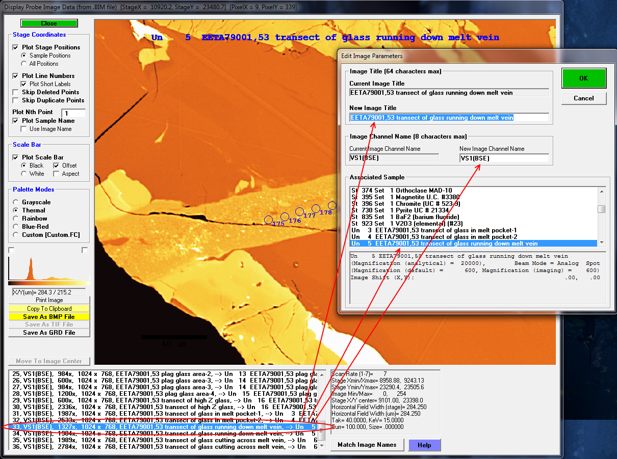
In addition, it should be noted that one can also "point to" any sample (and edit other fields) by simply double-clicking the image list item as seen here:


If you have any questions regarding this cool feature, please post them here.
« Last Edit: January 01, 2014, 09:27:56 PM by John Donovan »
John J. Donovan, Pres. 
(541) 343-3400

"Not Absolutely Certain, Yet Reliable"

John Donovan

  • Administrator
  • Emeritus
  • *****
  • Posts: 3304
  • Other duties as assigned...
    • Probe Software
Re: Correlating Images and Point Analyses in PFE
« Reply #1 on: June 30, 2014, 12:46:37 PM »
For best results, you'll want to re-calibrate your SE/BSE image calibration whenever your column is serviced and/or the beam scan or mag electronics are adjusted.

Start by clicking the "@" button in the Imaging window accessible from the Acquire! window as seen here (note that you need to have an image acquired to enable the button and the sample should be suitable for calibration purposes in the sense that it should contain at least two marks on the sample which the stage coordinates can be determined):


then, click "Yes" to continue as seen here:


then determine the stage coordinates and pixel coordinates by following the instructions as seen here:


Clicking the Calibrate button will provide one with the xmin/xmax and ymin/ymax stage coordinates that then need to be edited in the Probewin.ini file in the [image] section as seen here:

ImageInterfaceImageIxIy=1.3333      ; SX100 Video Ix/Iy (4:3 for SX100)
ImageInterfaceCalKeV=15.0      ; image calibration keV
ImageInterfaceCalMag=400.0      ; image calibration mag

;ImageInterfaceCalXMicrons=975.      ; SX100 Video X calibration in um (08-23-2010)
;ImageInterfaceCalYMicrons=700.      ; SX100 Video Y calibration in um (08-23-2010)
;ImageInterfaceCalXMicrons=948.      ; SX100 Video X calibration in um (08-23-2010)
;ImageInterfaceCalYMicrons=685.      ; SX100 Video Y calibration in um (08-23-2010)
;ImageInterfaceCalXMicrons=967.8   ; SX100 Video X calibration in um (09-21-2010)
;ImageInterfaceCalYMicrons=699.      ; SX100 Video Y calibration in um (09-21-2010)
;ImageInterfaceCalXMicrons=904.7   ; SX100 Video X calibration in um (09-23-2010)
;ImageInterfaceCalYMicrons=483.      ; SX100 Video Y calibration in um (09-23-2010)
;ImageInterfaceCalXMicrons=873.6   ; SX100 Video X calibration in um (09-23-2010)
;ImageInterfaceCalYMicrons=515.2   ; SX100 Video Y calibration in um (09-23-2010)
;ImageInterfaceCalXMicrons=875      ; SX100 Video X calibration in um (09-23-2010)
;ImageInterfaceCalYMicrons=521      ; SX100 Video Y calibration in um (09-23-2010)
ImageInterfaceCalXMicrons=943      ; SX100 Video X calibration in um (01-18-2011)
ImageInterfaceCalYMicrons=714      ; SX100 Video Y calibration in um (01-18-2011)

Note that only the last uncommented values for ImageInterfaceCalXMicrons and ImageInterfaceCalYMicrons are read by the application.  Be sure to edit the Probewin.ini values with the proper polarity (JEOL in reverse cartesian).

More information on this is here:

http://probesoftware.com/smf/index.php?topic=36.msg918#msg918
« Last Edit: June 30, 2014, 03:59:18 PM by John Donovan »
John J. Donovan, Pres. 
(541) 343-3400

"Not Absolutely Certain, Yet Reliable"

John Donovan

  • Administrator
  • Emeritus
  • *****
  • Posts: 3304
  • Other duties as assigned...
    • Probe Software
Re: Correlating Images and Point Analyses in PFE
« Reply #2 on: June 30, 2014, 12:54:55 PM »
By the way, one can easily compare the digitized positions with the acquired positions by acquiring an SE/BSE image after the acquisition has completed.

There are several things to keep in mind when looking at such images:

1. If the digitized positions were digitized using the Digitize Image feature, as described here:

http://probesoftware.com/smf/index.php?topic=70.msg263#msg263

and, if those positions are acquired using beam deflection, as described here:

http://probesoftware.com/smf/index.php?topic=70.msg266#msg266

then, the acquired positions will line up exactly with the saved digitized positions because the software uses the same image calibration to store the coordinates as it does to acquire them. Of course, if the stage has drifted since the points were acquired (e.g., beam was turned off and sample cooled down), then the post acquisition image spots where the sample was burned will not exactly line up with the beam deflection calibration.

The image calibration method is described here, but will be improved soon:

http://probesoftware.com/smf/index.php?topic=138.msg1429#msg1429

In the meantime, please keep in mind that if the sample positions were digitized using the Digitize Image feature and then subsequently acquired using stage motion, there may be some discrepency between the digitized and acquired positions depending on the accuracy of the image calibration.

The same applies to positions digitized using stage moves and subsequently acquired using beam deflection from the Automate! window.  Not to worry the application will warn you if the image magnification is too high to deflect the beam to all positions in each sample.

Here's an example where the beam deflection was utilized but the BSE image was acquired a day or so after the acquisition finished:

« Last Edit: June 30, 2014, 01:17:12 PM by John Donovan »
John J. Donovan, Pres. 
(541) 343-3400

"Not Absolutely Certain, Yet Reliable"

John Donovan

  • Administrator
  • Emeritus
  • *****
  • Posts: 3304
  • Other duties as assigned...
    • Probe Software
Re: Correlating Images and Point Analyses in PFE
« Reply #3 on: June 16, 2015, 05:44:32 PM »
For best results, you'll want to re-calibrate your SE/BSE image calibration whenever your column is serviced and/or the beam scan or mag electronics are adjusted.

For the most accurate beam scan calibration, you should utilize the new beam calibration method in CalcImage as described here:

http://probesoftware.com/smf/index.php?topic=396.0

It allows the user to quickly and automatically calibrate the beam scan X, Y extents and rotation for multiple magnifications and keVs by performing a mathematical transformation on over-lapping stage scan and beam scan images.

Very easy to use!
John J. Donovan, Pres. 
(541) 343-3400

"Not Absolutely Certain, Yet Reliable"

Gseward

  • Global Moderator
  • Professor
  • *****
  • Posts: 134
Re: Correlating Images and Point Analyses in PFE
« Reply #4 on: July 29, 2015, 01:38:33 PM »
John

Recently I noticed that the scale bar on my electron images is wrong. I checked my .ini  image mag calibrations and they seem to be ok. I opened the @ calibration tool in Imaging! and read the .ini values, but the calibration still seems to be off. Has something changed? or has it been so long since I looked at this that I'm missing something? Is it possible Cal Mag is not being read correctly, since it always says x400? Although since I manually entered the correct value in the .ini I don't see why this should matter.

Yes, I know I can now use CalcImage to do the calibration!

Cheers,


gareth
« Last Edit: July 29, 2015, 01:47:13 PM by Gseward »

Gseward

  • Global Moderator
  • Professor
  • *****
  • Posts: 134
Re: Correlating Images and Point Analyses in PFE
« Reply #5 on: July 29, 2015, 01:52:03 PM »
John.

I fixed it. Even though I didn't change the values in the .ini, I ran though the calibration procedure and now the correct values are being used. It seems simply clicking 'reload .ini values' was not reloading them?
gareth

Probeman

  • Emeritus
  • *****
  • Posts: 2856
  • Never sleeps...
    • John Donovan
Re: Correlating Images and Point Analyses in PFE
« Reply #6 on: July 29, 2015, 02:41:23 PM »
John.

I fixed it. Even though I didn't change the values in the .ini, I ran though the calibration procedure and now the correct values are being used. It seems simply clicking 'reload .ini values' was not reloading them?
gareth

Hi Gareth,
I don't quite understand what may have happened.  But just for the record, the "ReLoad INI values" button is just there as a convenience, to avoid forcing the user close and re-open PFE to load new values after editing the probewin.ini file for new calibrations.

That said, the @ button should now only be used for quick and dirty image calibrations or maybe just to get started.  This new method using three points which automatically calibrates X, Y and image rotation is described here should be used from now on:

http://probesoftware.com/smf/index.php?topic=396.0

And it will edit your Probewin.ini file automatically. And it works for Probe Image calibrations and also Thermo or Bruker image calibrations (though it won't edit the stored calibrations automatically as it will in PFE).

I would be very interested in someone trying this new method and posting some results here.
The only stupid question is the one not asked!

Gseward

  • Global Moderator
  • Professor
  • *****
  • Posts: 134
Re: Correlating Images and Point Analyses in PFE
« Reply #7 on: July 30, 2015, 12:28:19 PM »
John,

I think I found the cause of the issue:

If 'Do Not Set Conditions During Acquisition' is selected from the Acquisition Options window, then images that are collected do not have the correct spatial registration. See example below.

Is this perhaps because the mag is not being read? It seems that even though conditions are not being set, you probably intended for them to be read, right?

In these screen snaps you can see the same image acquired twice, first with 'Do Not Set Conditions During Acquisition' selected, and then with it deselected. Note that the mag is the same, and the same acquired analysis points have  been overlaid (incorrectly in the first image, as shown by the contamination marks in the SE image (green circles)).


Incorrect overlay


Correct overlay

John Donovan

  • Administrator
  • Emeritus
  • *****
  • Posts: 3304
  • Other duties as assigned...
    • Probe Software
Re: Correlating Images and Point Analyses in PFE
« Reply #8 on: July 30, 2015, 01:41:46 PM »
If 'Do Not Set Conditions During Acquisition' is selected from the Acquisition Options window, then images that are collected do not have the correct spatial registration. See example below.

Is this perhaps because the mag is not being read? It seems that even though conditions are not being set, you probably intended for them to be read, right?

Nice catch! 

I don't think it is due to not reading the mag since the mag displayed is the same for both images, but yes, I'm skipping something that I shouldn't be when the "Do Not Set Conditions Before Acquisition" option is checked.
john
John J. Donovan, Pres. 
(541) 343-3400

"Not Absolutely Certain, Yet Reliable"

John Donovan

  • Administrator
  • Emeritus
  • *****
  • Posts: 3304
  • Other duties as assigned...
    • Probe Software
Re: Correlating Images and Point Analyses in PFE
« Reply #9 on: July 31, 2015, 12:21:48 AM »
If 'Do Not Set Conditions During Acquisition' is selected from the Acquisition Options window, then images that are collected do not have the correct spatial registration. See example below.

Is this perhaps because the mag is not being read? It seems that even though conditions are not being set, you probably intended for them to be read, right?

Nice catch! 

I don't think it is due to not reading the mag since the mag displayed is the same for both images, but yes, I'm skipping something that I shouldn't be when the "Do Not Set Conditions Before Acquisition" option is checked.
john

Fixed and ready to download now.
John J. Donovan, Pres. 
(541) 343-3400

"Not Absolutely Certain, Yet Reliable"