Hi Karen,
Great question!
Probe Image and CalcImage use all the background measurement and modeling options used in Probe for EPMA with the exception of the multi-point background method. For a comparison of off-peak, MAN and Nth point background methods see this post:
http://probesoftware.com/smf/index.php?topic=4.msg189#msg189So for example, you can run x-ray maps using an MAN calibration curve which is good to 100-200 PPM in oxides and silicates. For higher Z materials there are some work that has been done at GE and University of Tasmania. See this thread for more info:
http://probesoftware.com/smf/index.php?topic=4.msg17#msg17The advantage of MAN background corrections for x-ray mapping of course is that it cuts your analysis time in half or more. I've attached some nice images that illustrate this type of background correction for quantitative x-ray mapping. Also please see this thread on a new technique I've found using MAN bgds and the "blank correction" to get excellent accuracy, improve precision and save time all together! But only if your unknown has a simple enough matrix that one can produce a suitable "blank" standard material such as SiO2, TiO2, ZrSiO4, etc. See this thread for more details:
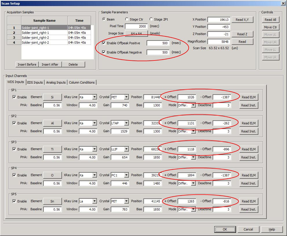
http://probesoftware.com/smf/index.php?topic=29.msg237#msg237In addition, in Probe Image one can acquire a single off-peak (high or low offset) and also high *and* low offset images automatically, as seen here in this screenshot of Probe Image where one can see that because Probe Image reads the background offsets as specified by the user in the Probe for EPMA run (containing the standard intensities) you only need to check or uncheck these background checkboxes as desired.
Of course, just as in Probe for EPMA, once you are quantifying x-ray maps in CalcImage, you get full access to all the cool off-peak modeling options in PFE (with the one exception already noted above for multi-point bgds)!


 If you can't see some "in-line" images in a post, try "refreshing" your browser!